Implant Osseointegration Protocol
Sectioning and Staining
Click image to enlarge.
Osseointegration normally relies on ground sections because it’s generally not possible to cut metal implants on a microtome. Surface staining methods are therefore required.
Bone Formation Boundary
Click image to enlarge.
A standard technique in osseointegration is the analysis of new bone formation and bone contact surface within a standard zone around the implant. BIOQUANT includes tools to standardize the thickness of the sampling area around the implant. A zone thickness is specified and then a drawing tool guides a technician in tracing out the sampling area. In the image at the top, a zone thickness of 125 microns was used.
Referent Data
Click image to enlarge.
Once the sampling area is defined, BIOQUANT automatically collects three pieces of referent data used to normalized the other values measured on the section:
Im.Ar (Implant Area)
Im.Pm (Implant Perimeter)
Sa.Ar (Sampling Area)
FYI - The Referent Data Tool can be used on a large scan of the tissue, zoomed out to fit in the Image window as show above. It can also be used field-to-field at higher magnification. You do not need a scan or tracking device to use BIOQUANT, although they do save you time.
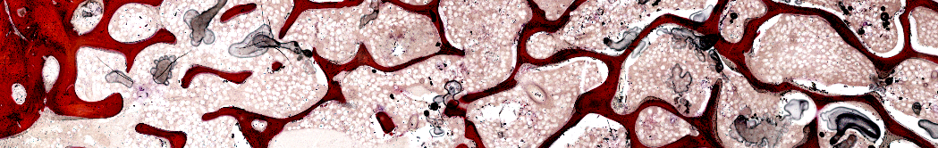
Bone Structure
Click image to enlarge.
Referent Data Tool
Bone in the sampling is identified by specific staining and color-based thresholding tools. Draw and Erase tools allow corrections to be made to the automatic threshold. Once the bone is defined, BIOQUANT measures:
B.Ar, B.Pm
Tb.N, Tb.Sp, Tb.Dm
Bone Contact Surface
Click image to enlarge.
The portion of the implant surface that is occupied by bone is marked at high magnification. BIOQUANT then computes:
Co.Pm
Co.Pm/B.Pm
Co.Pm/Im.Pm
Repeat for Remaining Implant Surface
Click image to enlarge.
Recent Citations of BIOQUANT in Implant Research
Browse additional citations of BIOQUANT in implant research...