
Muscle Fiber Phenotyping
Multi-channel Fluorescence Imaging
Multi-channel fluorescent staining indicating fibertype. Click to enlarge.
For multi-channel fluorescent labeling, it is recommended to use a multi-band fluorescent filter cube and a color digital camera for imaging. The relative intensity of labels can be adjusted within the camera controls.
This speeds up imaging since you only have to capture one image per field of view and you don't have to apply false coloring to monochrome images and the merge them to create the same color image.
Defining the Muscle Boundary
At low magnification, the boundary of the muscle is traced using the irregular ROI (region of interest) tool. This allows BIOQUANT to count only myofibers within the muscle when working at higher magnification.
Automated Myofiber Detection
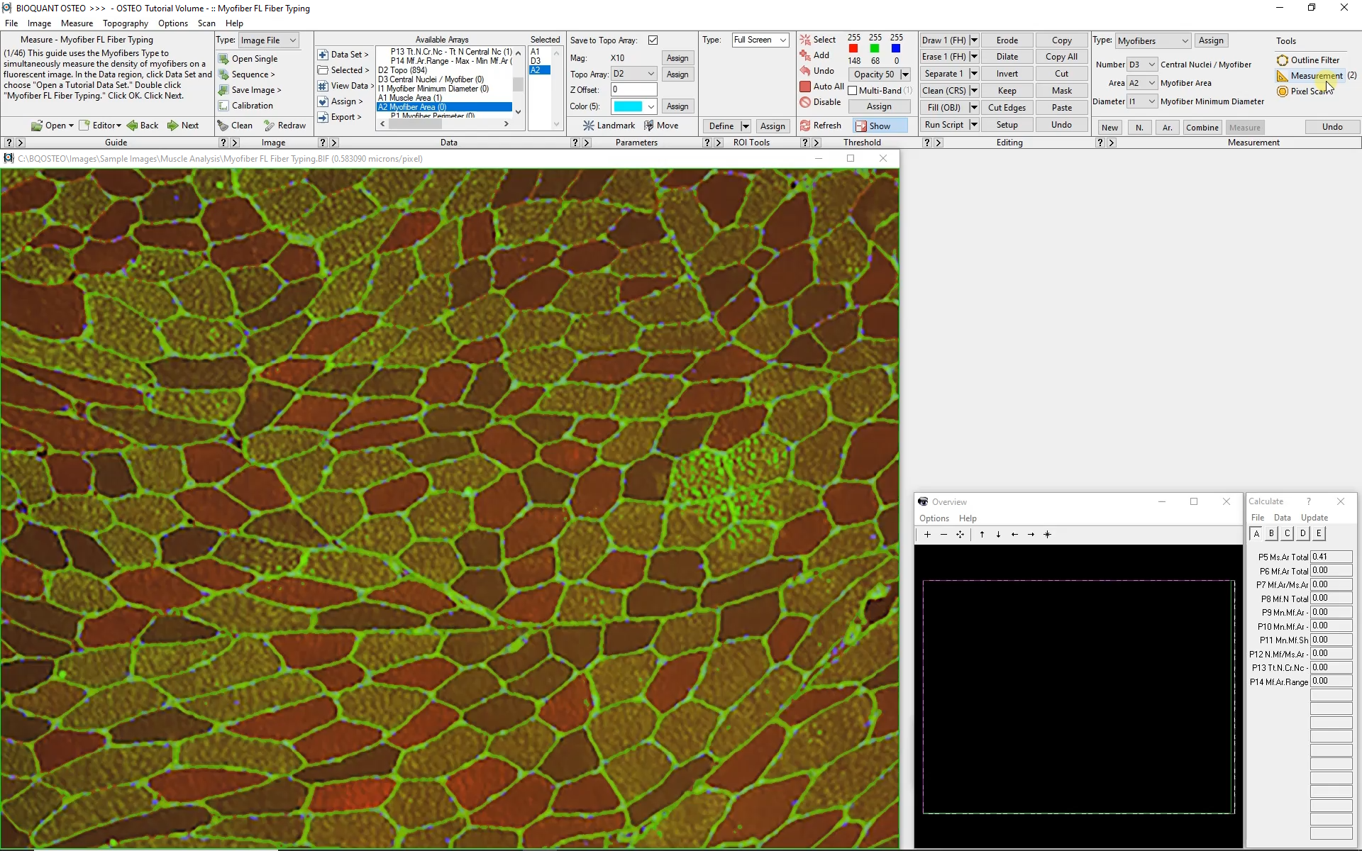
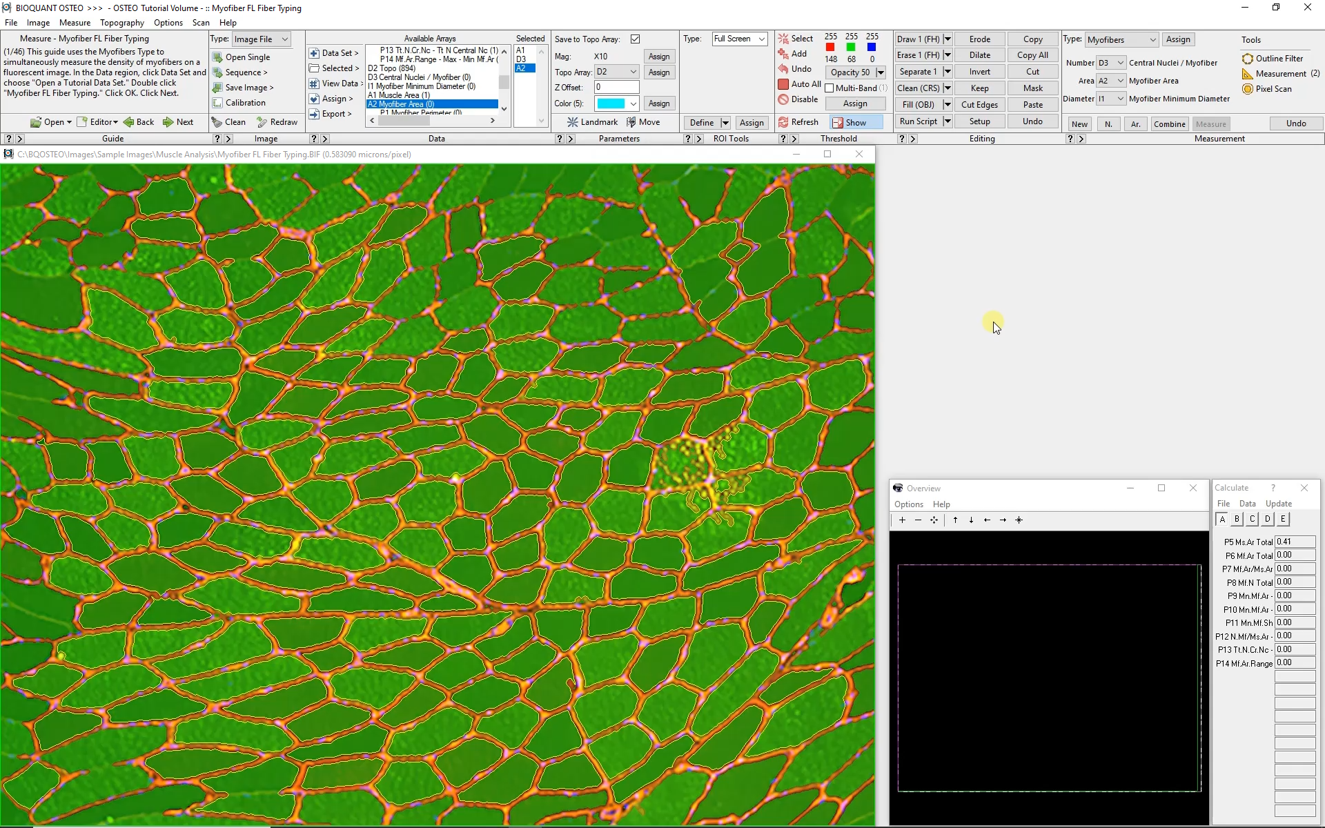
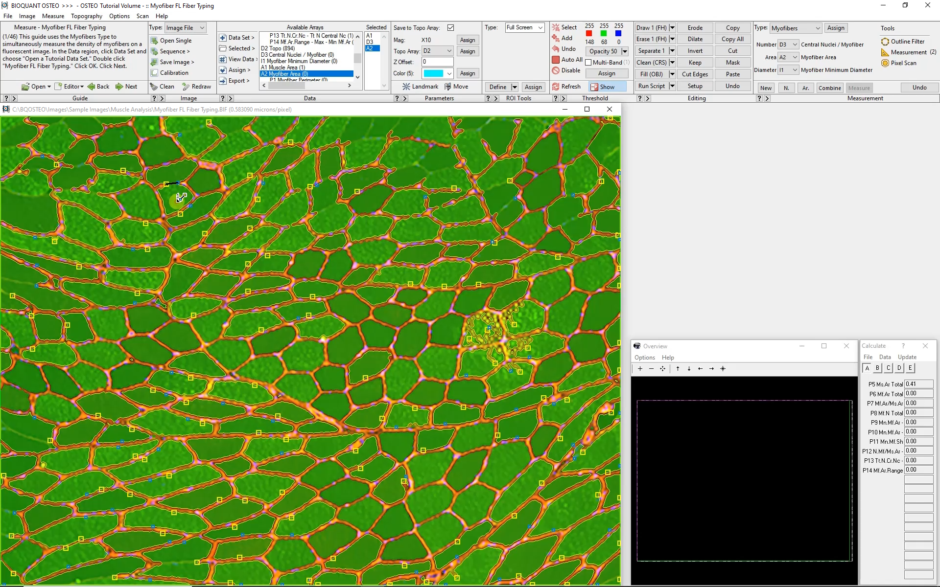
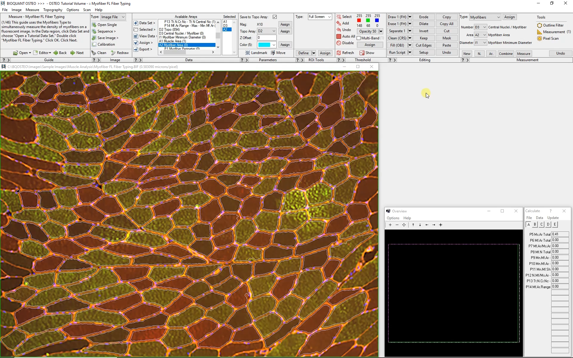
Automated analysis of myofibers depends on a high-contrast label applied to the boundary of each myofiber. Generally this requires a label specific to dystrophin or laminin.
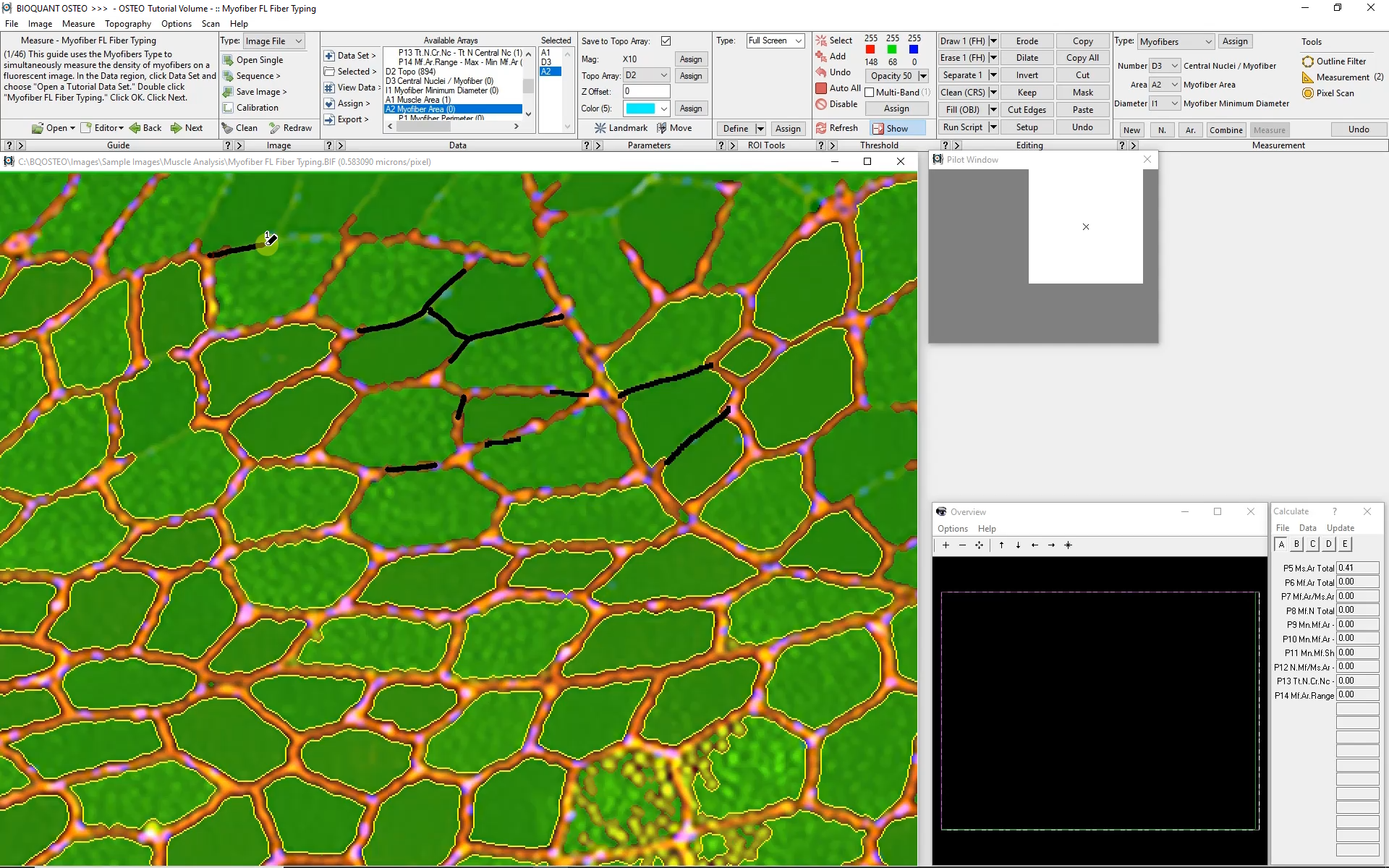
Automatic color thresholding uses this boundary stain to identify the myofibers. Manual editing with a brush and eraser makes it simple to correct mistakes.
Intelligent filters remove previously measured myofibers and myofibers that are not entirely visible within the field of view.
Measuring Myofibers
Click to Enlarge Image
BIOQUANT simultaneously collects the following data from each myofiber:
Myofiber Cross-sectional Area
Myofiber Shortest Diameter
Myofiber Perimeter
Myofiber Location
Myofiber Circularity
Myofiber Fibertype
Number of Myofibers
Number of Central Nuclei per Myofiber
Subsequent Fields of View
The Large Image Navigator in BIOQUANT makes it simple to move sequentially through a large section in consecutive, overlapping fields of view. BIOQUANT automatically tracks the boundary of the muscle and skips fields of view which are outside the muscle boundary.