
Axon Number and Caliber
Toluidine Blue Imaging

In Glaucoma and Neuropathy Models
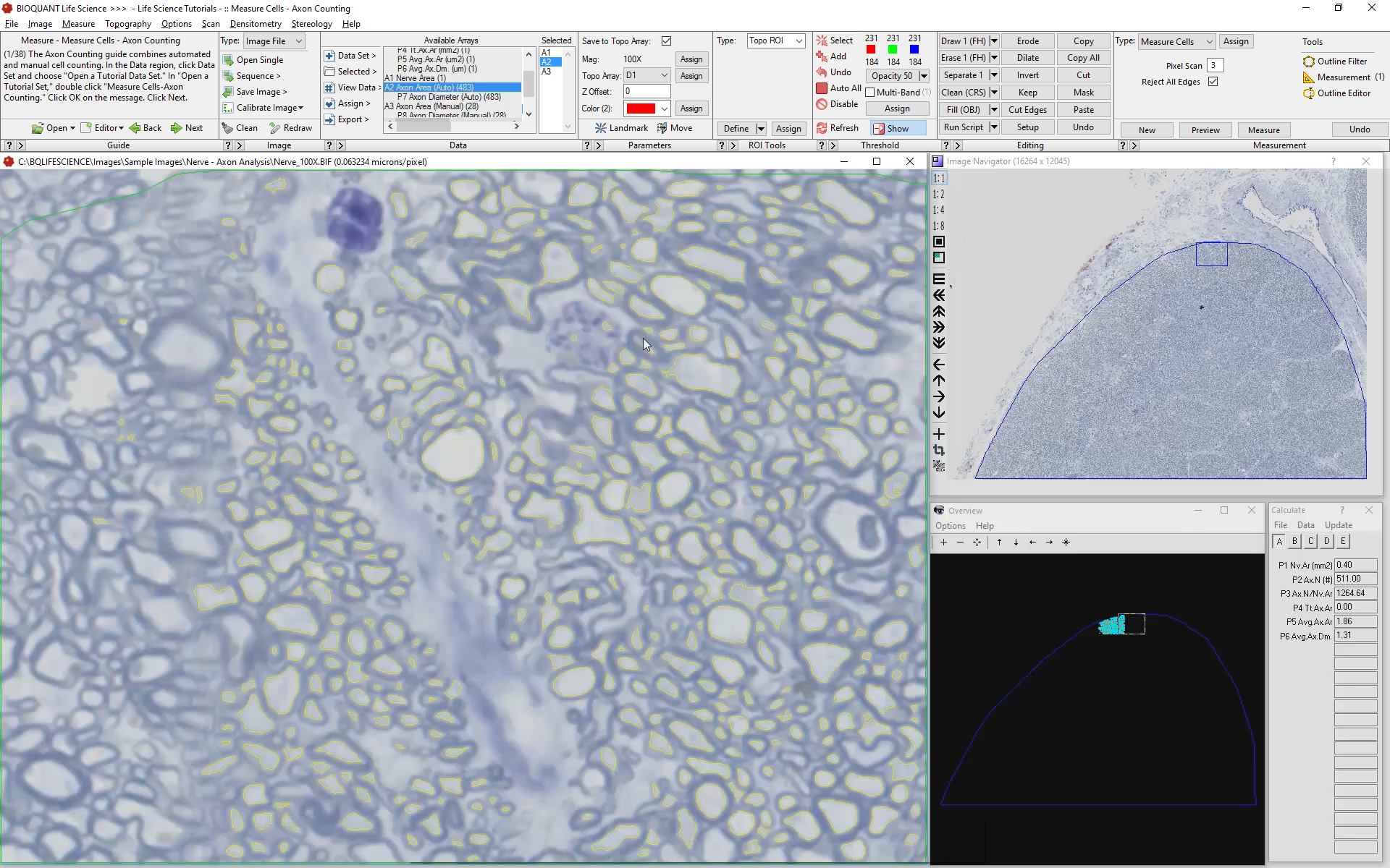
In semi-thin sections with myelin specific staining, BIOQUANT automatically detects myelinated axons. Detection is based on color, shape, and size. Shape filters automatically exclude obliquely cut axons.
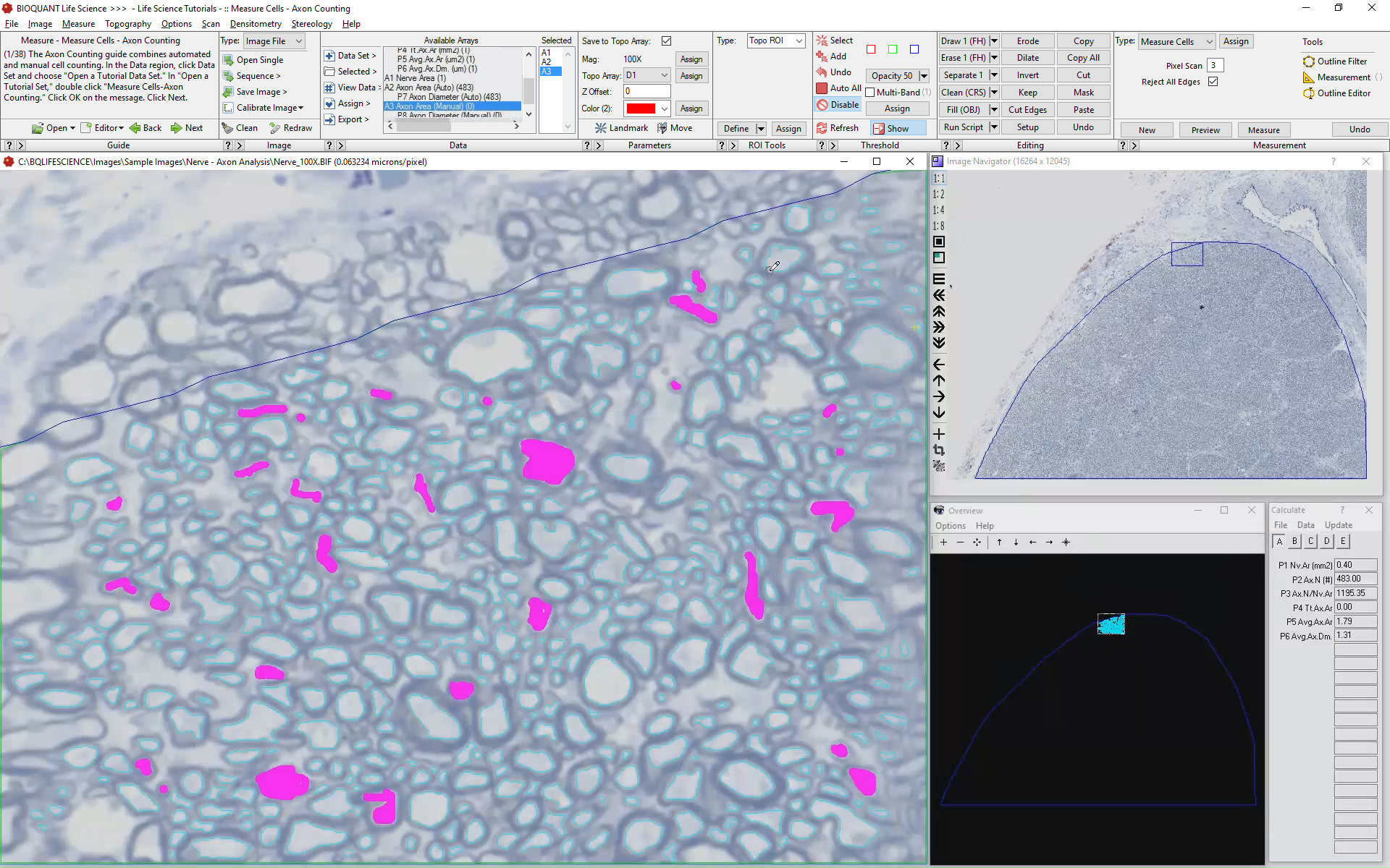
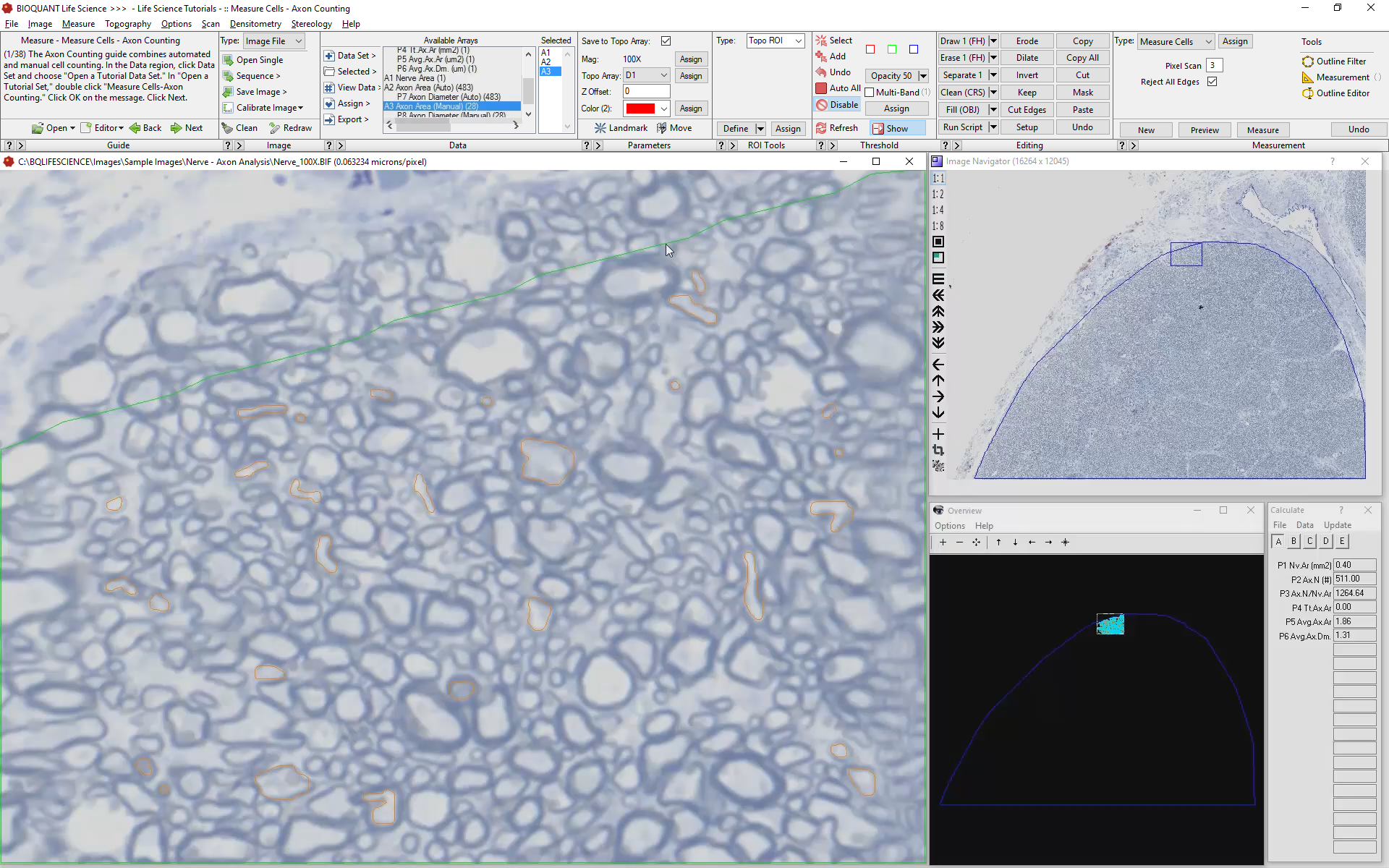
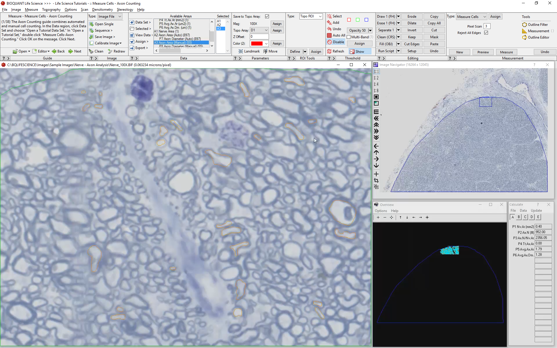
In the case of very small axons or partially myelnated axons, some manual editing may be necessary.
A unique object recognition algorithm prevents the system from double-counting when scanning through adjacent overlapping fields of view.
Large scale projects have counted 50% of the axons in a non-human primate optic nerve.
Define and Collect Nerve Data
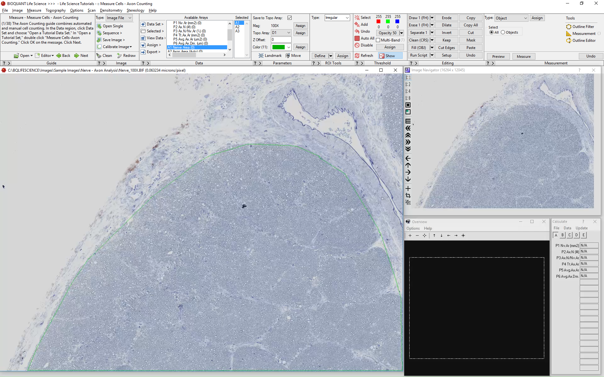
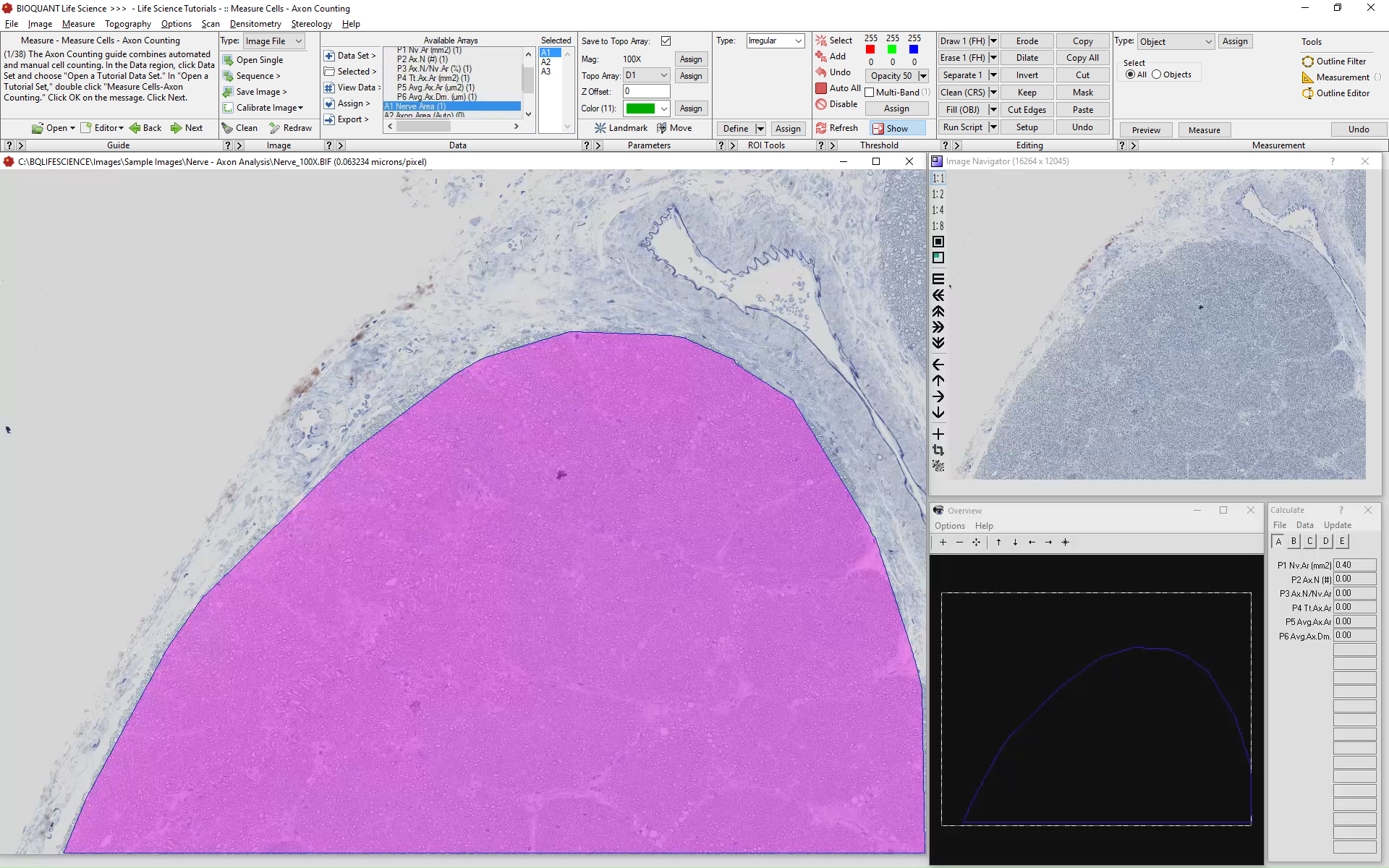
Define the nerve borders using the Irregular Region of Interest tool. BIOQUANT will limit threshold to within the ROI and retrieve Nerve Area data.
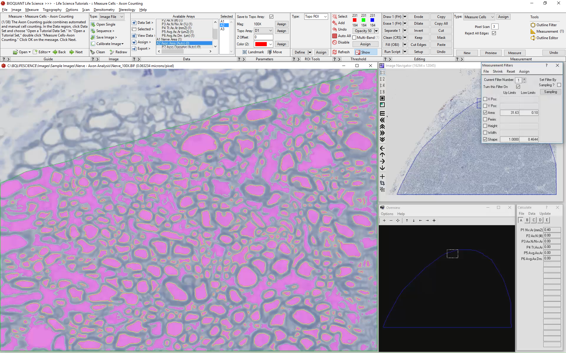
Collect Axon Data
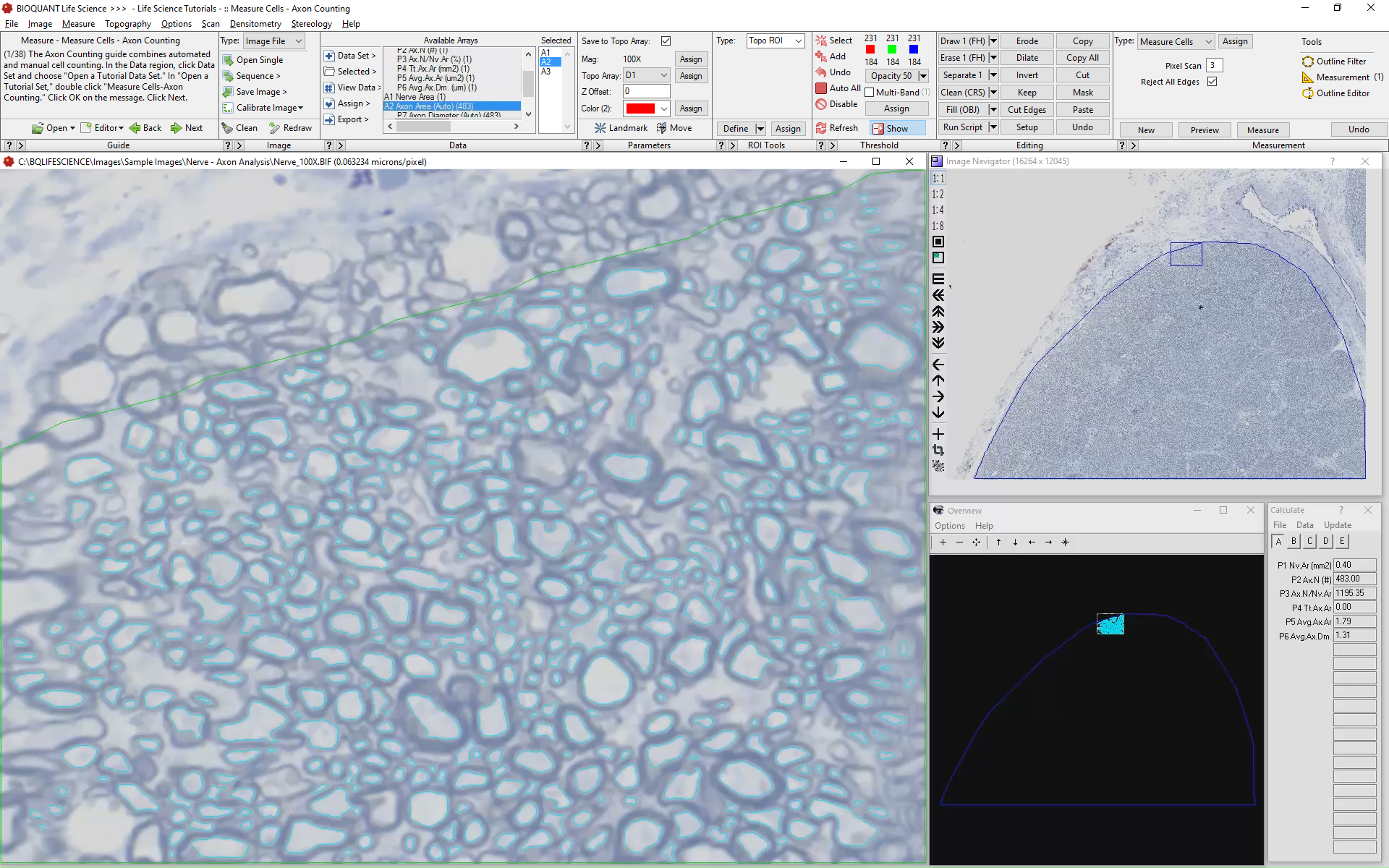
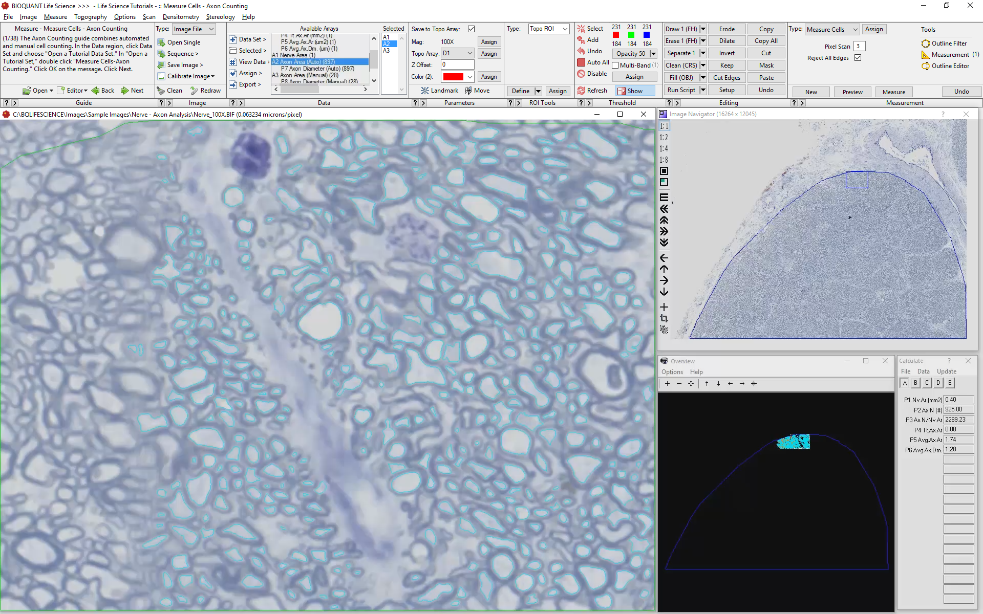
Based on stain color BIOQUANT will threshold the individual axons.

BIOQUANT then automatically employs Measurement Filters based on axon area and shape. The histologist will remove false positives using BIOQUANTs Editing Tools, then Measure the current field of view.
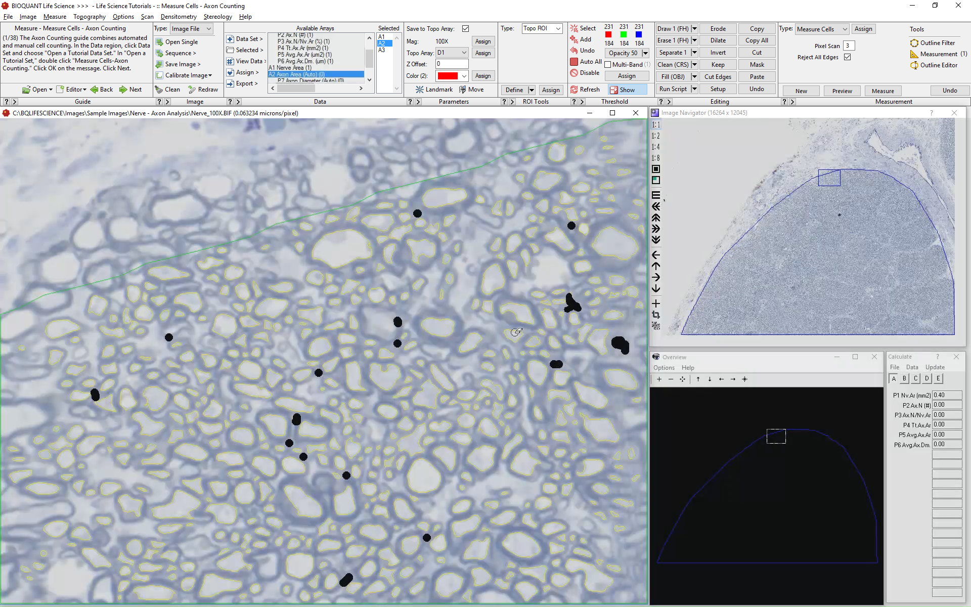
Lastly, the histologist will add axons that did not threshold, or remove false positive axons using BIOQUANTs Editing Tools.
Move to the Next Field of View
Once done with this field of view, BIOQUANT will sequence to the next view, then repeat the protocol. BIOQUANT automatically rejects axons touching the edge of the screen and previously measured axons.


Computed Data
Nerve Area
Individual Axon Area
Individual Axon Caliber
Axon Count
Axon Density (#/unit area)
Mean Axon Area
Mean Axon Caliber