Retina Layer Structure
Image with Toluidine Blue
TolBlue is an excellent marker for each layer in a retinal cross section.

Cell Number and Layer Thicknesses
In retinal cross-sections, a specialized tool analyzes the thickness of consecutive layers.
Once the boundaries between layers are traced, perpendicular thickness is measured automatically at regularly spaced intervals along the surface.
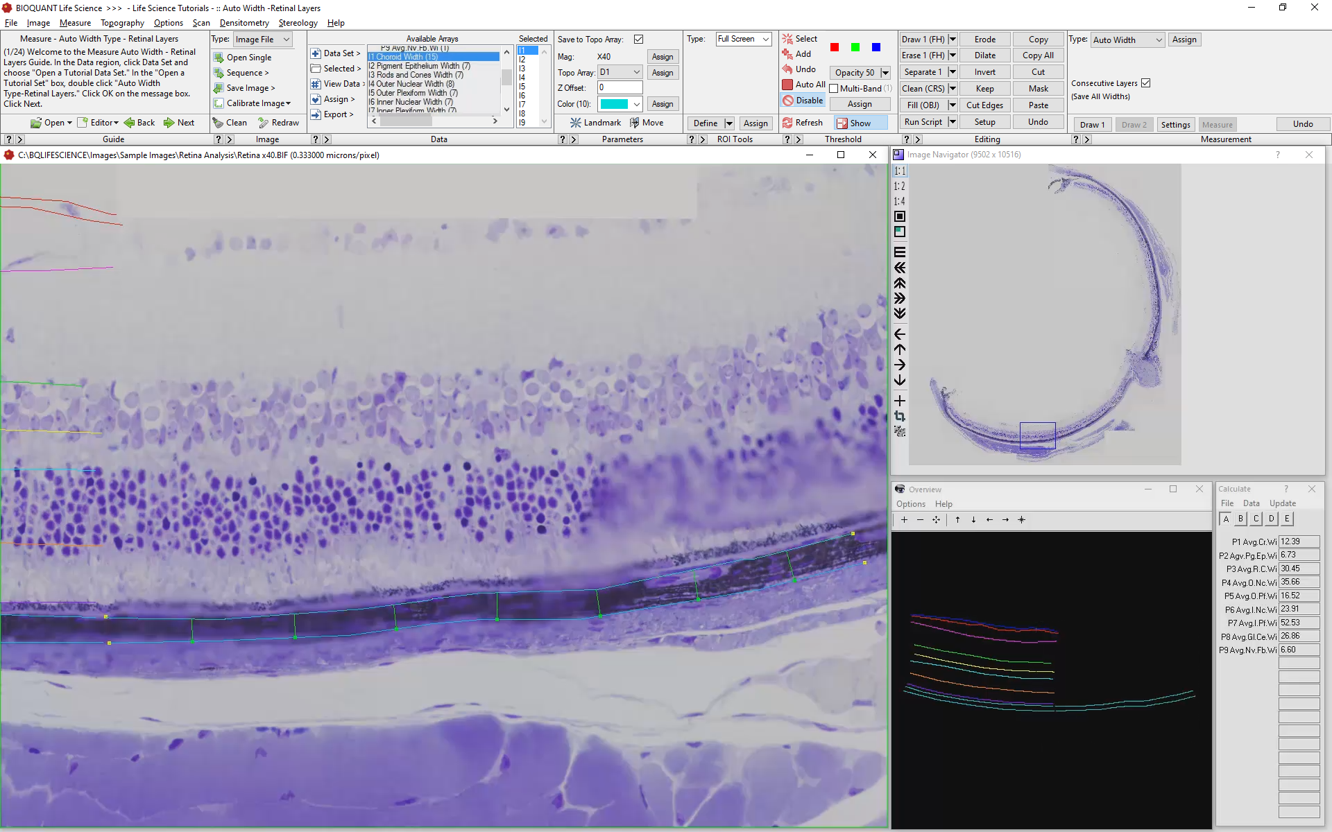
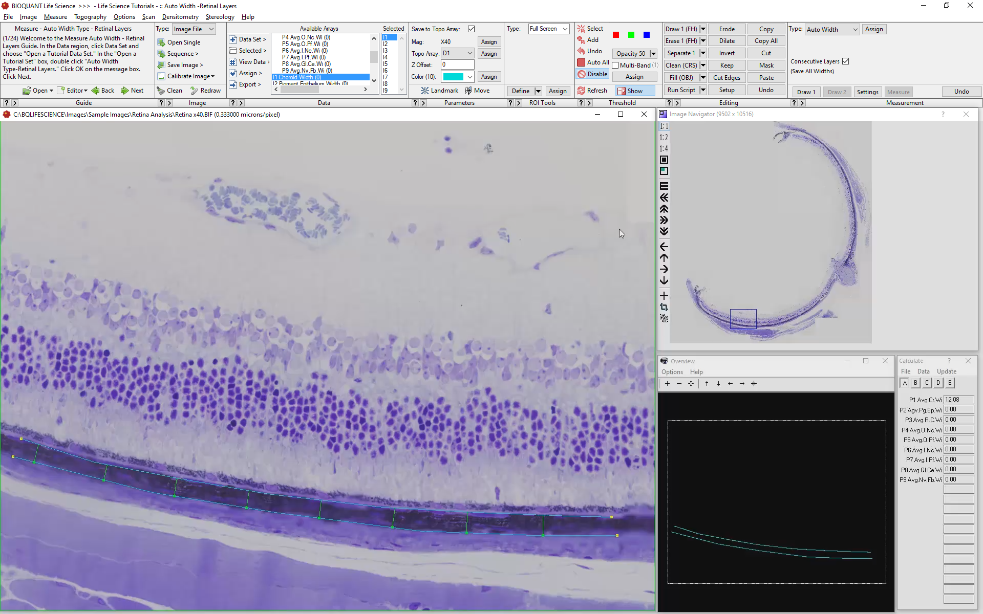
Collect Choroid Layer Width Data
Using BIOQUANTs Auto Width tool, first draw the outer, then inner surfaces of the choroid layer. BIOQUANT will automatically generate perpendicular, unbiased lines.

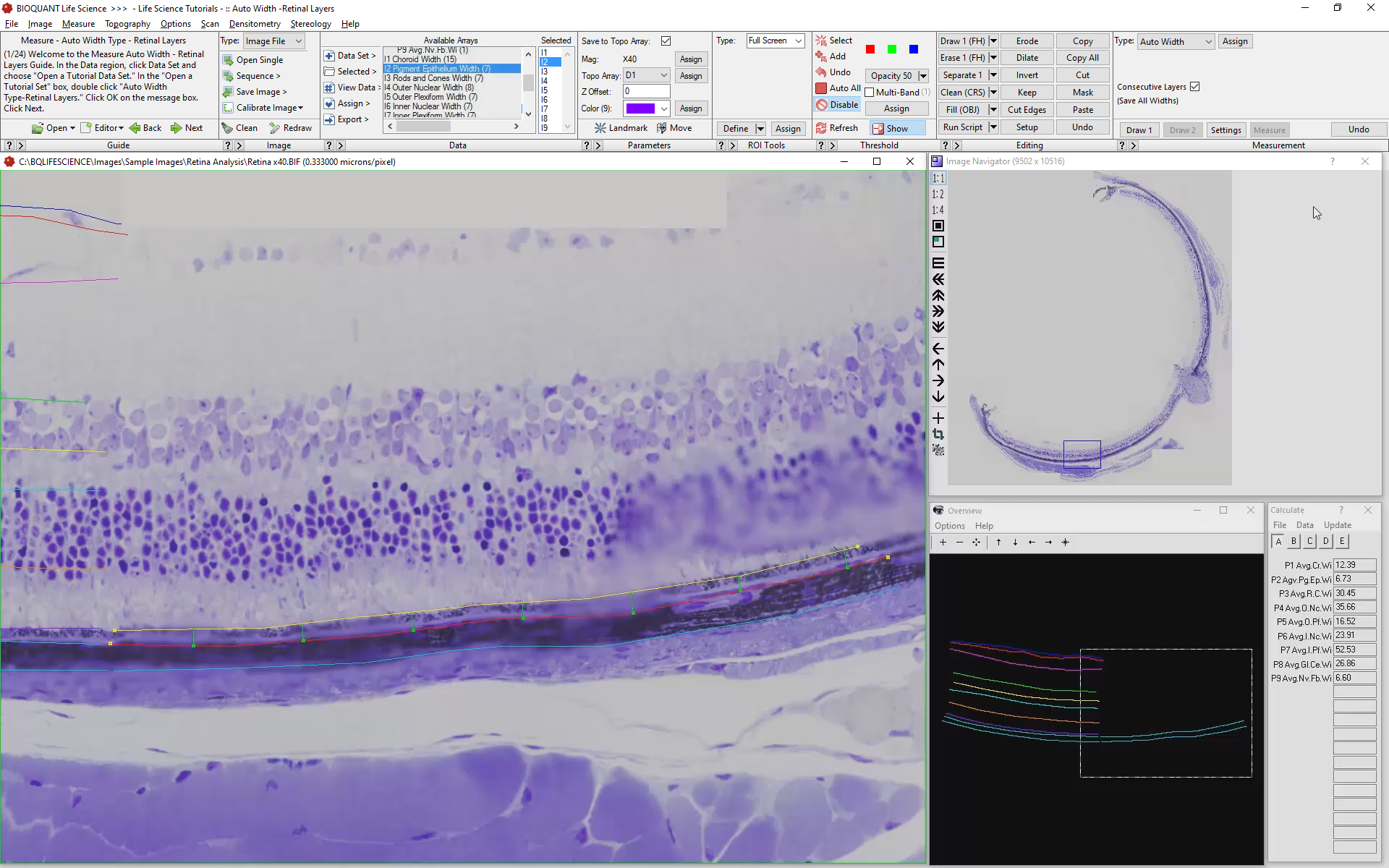
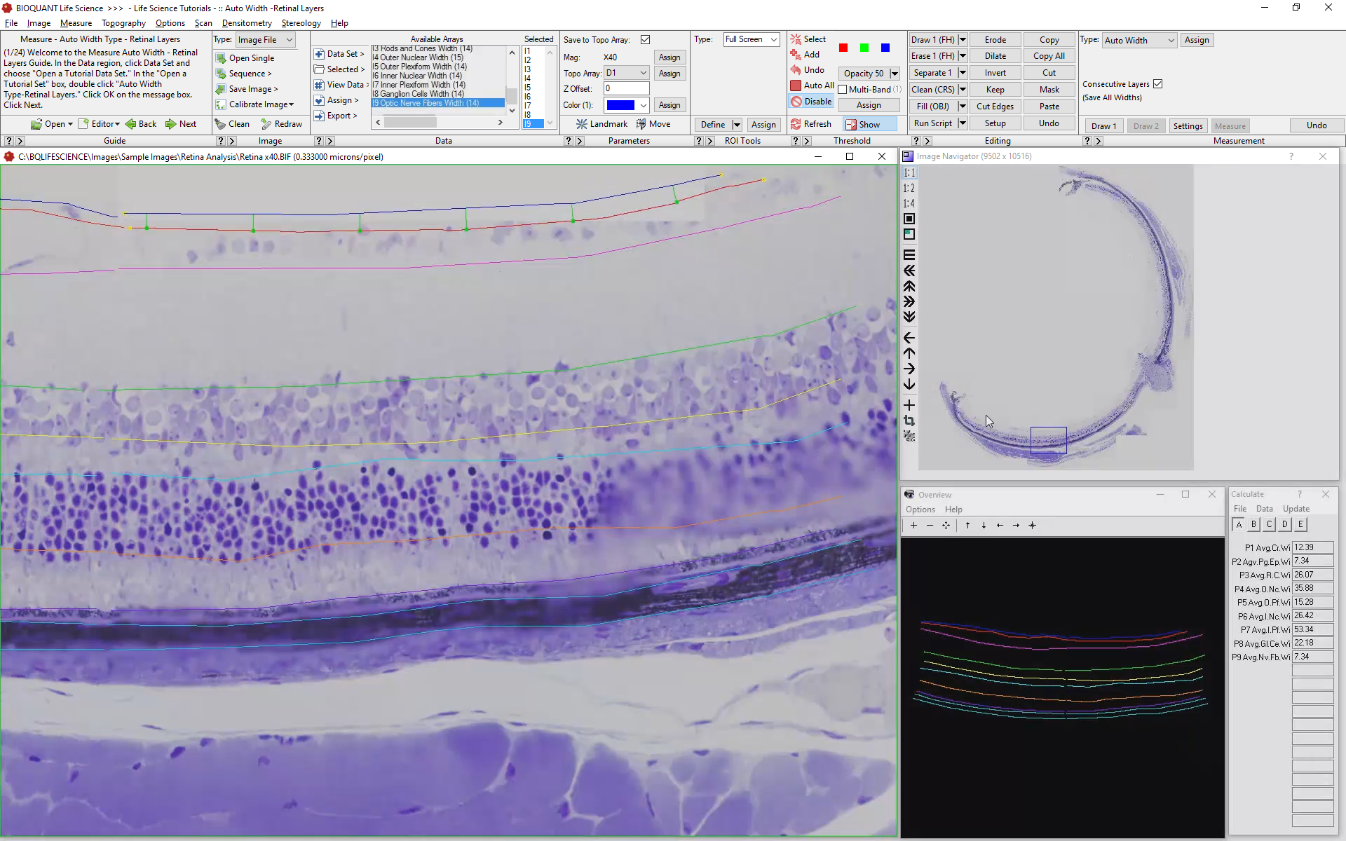
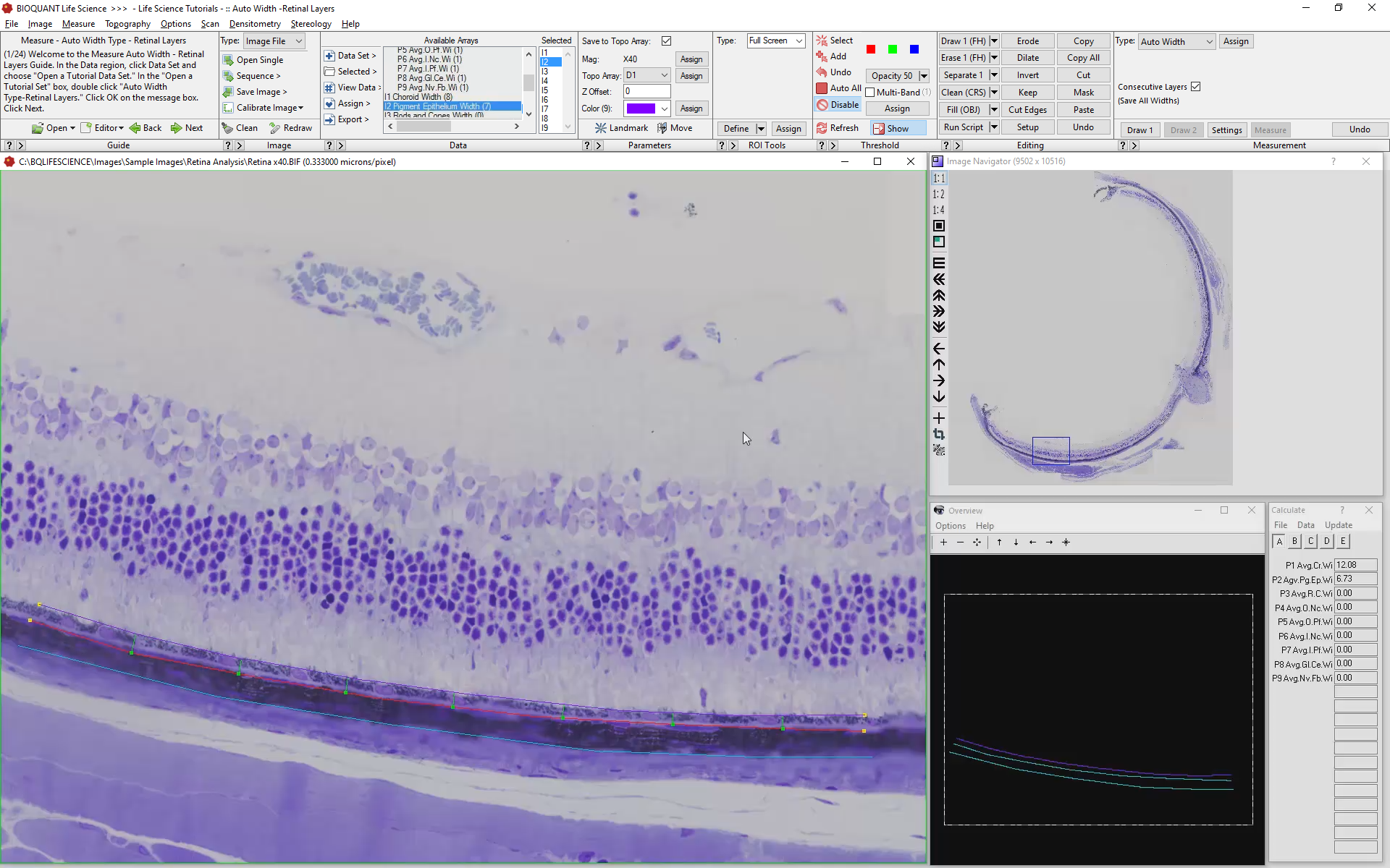
Draw Each Sequential Layer
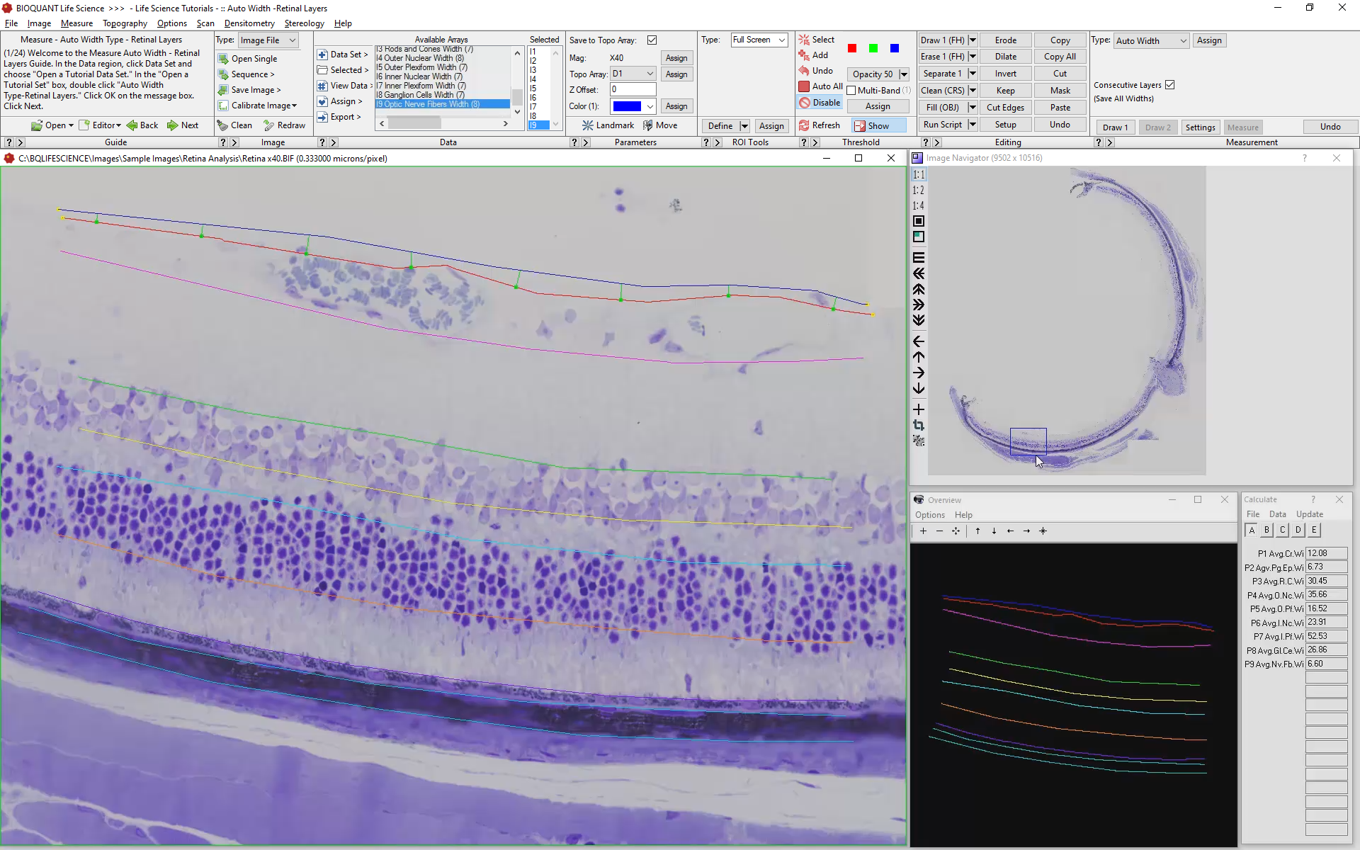
Because BIOQUANT already knows where you left off with each layer, simply draw the inner surface of the new layers, measuring in between. For each field of view you will get a running total for:
Choroid Width
Pigment Epithelium Width
Rod /Cone Width
Outer Nuclear Width
Outer Plexiform Width
Inner Nuclear Width
Inner Plexiform Width
Ganglion Cell Width

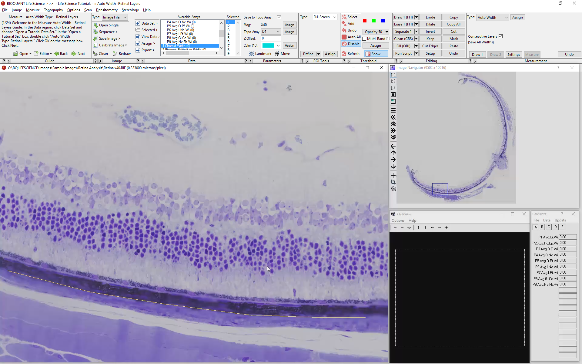
Move to the Next Field of View
On an overlapping field of view, you can see exactly where you left off. You may even leave for the day and continue the next.

Repeat the Protocol
Simply repeat the protocol for each remaining field of view. BIOQUANT will keep track of individual widths, showing you the running average in the calculations box.